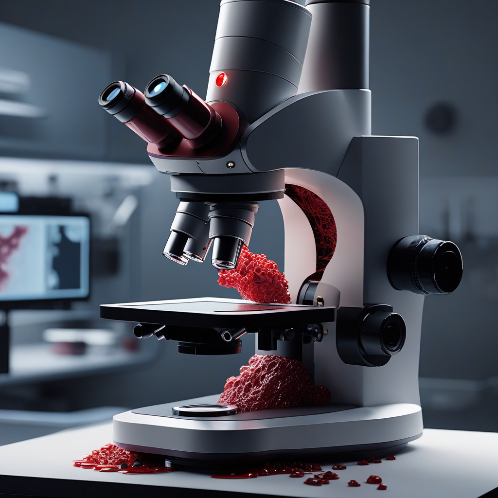
AI Microscope Revolutionizes Blood Clot Detection by Observing Live Platelet Clumping Instantly
In a groundbreaking development in the field of medical technology, scientists have successfully utilized artificial intelligence (AI) in microscopes to detect live blood clot activity. This innovation allows researchers to observe platelet clumping in real time, providing valuable insights into the mechanisms of blood clot formation and potential treatment strategies.
Traditionally, studying blood clotting processes involved time-consuming and labor-intensive techniques that often provided limited information. However, with the integration of AI technology into microscopes, scientists can now monitor and analyze platelet behavior with unprecedented speed and accuracy.
One of the key advantages of this AI-powered microscope is its ability to capture dynamic changes in platelet activity at the cellular level. By leveraging machine learning algorithms, the system can identify and track individual platelets in real time, offering a comprehensive view of how these blood cells interact and aggregate to form clots.
Moreover, the high-resolution imaging capabilities of the AI microscope enable researchers to visualize the intricate details of clot formation, such as the speed and extent of platelet aggregation. This level of precision not only enhances our understanding of the clotting process but also opens up new possibilities for targeted interventions to prevent or treat abnormal clotting disorders.
The implications of this technological advancement extend beyond basic research, as it holds great promise for clinical applications as well. For instance, by detecting abnormal clotting patterns early on, healthcare providers can intervene proactively to prevent potentially life-threatening conditions such as heart attacks or strokes.
Furthermore, the real-time monitoring capabilities of the AI microscope could revolutionize the field of personalized medicine by allowing for tailored treatment approaches based on an individual’s unique clotting profile. This level of customization has the potential to improve patient outcomes and reduce the risk of complications associated with conventional, one-size-fits-all treatment strategies.
As with any innovation, there are still challenges to overcome in the widespread adoption of AI microscopes for blood clot detection. Issues such as standardization of imaging protocols, data interpretation, and integration with existing diagnostic workflows will need to be addressed to ensure seamless implementation in research and clinical settings.
Nevertheless, the successful demonstration of AI-powered microscopy for observing live blood clot activity represents a significant step forward in our ability to unravel the complexities of hemostasis and thrombosis. By harnessing the power of artificial intelligence, scientists and healthcare professionals are poised to make transformative advancements in the diagnosis and management of clotting disorders.
In conclusion, the integration of AI technology into microscopes has the potential to revolutionize the field of blood clot detection by enabling real-time observation of platelet clumping. This innovation not only enhances our understanding of clotting processes at the cellular level but also paves the way for personalized treatment strategies that could improve patient outcomes significantly.
AI microscope, Blood clot detection, Platelet clumping, Medical technology, Machine learning algorithms